
Simple Limbal Epithelial Transplantation (SLET) in a patient with limbal stem cell deficiency
Dilek Dursun Altınors1, Leyla Asena1, Sibel Oto1.
1Ophthalmology, Baskent University Faculty of Medicine, Ankara, Turkey
Introduction: When corneal epithelial stem cells become dysfunctional or deficient, limbal stem cell deficiency (LSCD) develops.1 LSCD leads to neovascularization of the corneal surface, persistent epithelial defects, scarring and decreased vision. Simple limbal epithelial transplantation (SLET) is a relatively new technique for addressing limbal stem cell deficiency.2 Limbal tissue from the patient's fellow healthy eye, is transplanted onto an amniotic membrane attached to the surface of the diseased eye, where they can expand in vivo.3 Here, we describe a case with unilateral limbal stem cell deficiency who was treated with simple limbal epithelial transplantation.
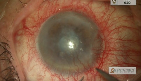
Case: A 78-year-old male patient admitted to our clinic with long standing herpetic stromal keratitis in his left eye. He was treated with systemic and topical antiviral medication and topical corticosteroids. At the end of the medical treatment period, the inflammation was inactive however there was a vascularized pannus covering the entire corneal periphery indicating corneal limbal stem cell deficiency, resulting in decreased visual acuity (Figure 1). The patient was diagnosed as LSCD secondary to herpetic neurotrophic keratitis and SLET from the healthy contralateral eye was performed.
Figure1: Preoperative anterior segment photograph.
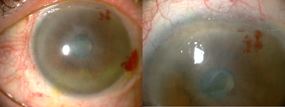
A 2 × 2 mm strip of donor limbal tissue was obtained from the healthy eye and divided into eight to ten small pieces. After surgical preparation of the recipient ocular surface by superficial keratectomy and pannus excision, these tiny limbal transplants were distributed evenly and attached using fibrin tissue adhesive over an amniotic membrane placed on the cornea (Figure 2).
Figure 2: Postoperative first day anterior segment photograph.
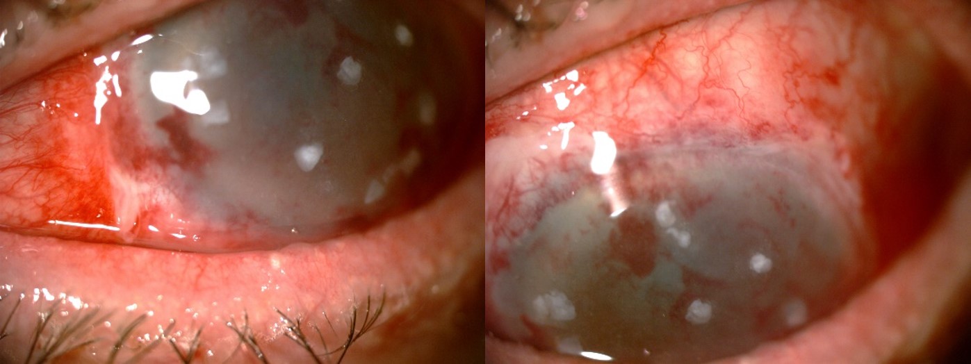
At the fourth postoperative week a completely epithelialized, avascular and stable corneal surface was observed (Figure 3), and this was maintained during a follow-up of 9 months (Figure 4). Best corrected Snellen visual acuity improved from 0.5 to 0.9.
Figure 3: Postoperative first month anterior segment photograph.
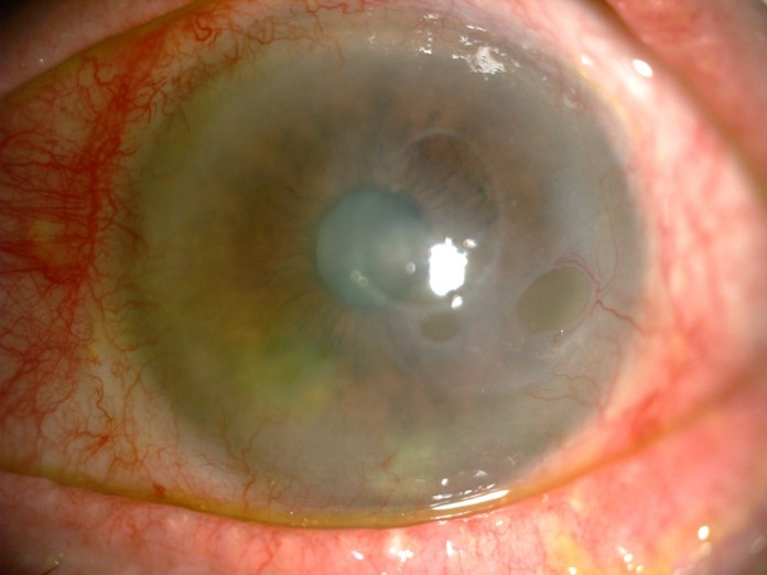
Figure 4: Postoperative nine months anterior segment photograph.
Discussion: SLET is a relatively new surgical technique for limbal stem cell transplantation which requires less donor tissue than previously used for conventional autografting and does not need a specialist laboratory for cell expansion. A single-center analysis of 125 cases of autologous SLET for unilateral LSCD demonstrated a 76% success rate and a 75% two-line improvement in visual acuity with a median follow-up of 1.5 years.4 Similarly, a multi-center analysis of 68 cases of autologous SLET for unilateral LSCD demonstrated an 84% success rate and a 65% two-line improvement in visual acuity with a median follow-up of one year.5
Conclusion: SLET is an easy and effective technique for treating unilateral LSCD which seems a highly successful surgical technique in mild to moderate LSCD.
[1] Holland EJ. Management of limbal stem cell deficiency: A historical perspective, past, present, and future. Cornea. 2015;34(Suppl 10):S9–15.
[2] Sangwan VS, Sharp JAH. Simple limbal epithelial transplantation. Curr Opin Ophthalmol. 2017 Jul;28(4):382-386.
[3] Yin J, Jurkunas U. Limbal Stem Cell Transplantation and Complications. Semin Ophthalmol. 2018;33(1):134-141.
[4] Basu S, Sureka SP, Shanbhag SS, Kethiri AR, Singh V, Sangwan VS. Simple limbal epithelial transplantation: Long-term clinical outcomes in 125 cases of unilateral chronic ocular surface burns. Ophthalmology. 2016;123 (5):1000–1010.
[5] Vazirani J, Ali MH, Sharma N, et al. Autologous simple limbal epithelial transplantation for unilateral limbal stem cell deficiency: Multicentre results. Br J Ophthalmol. 2016;100 (10):1416–1420.